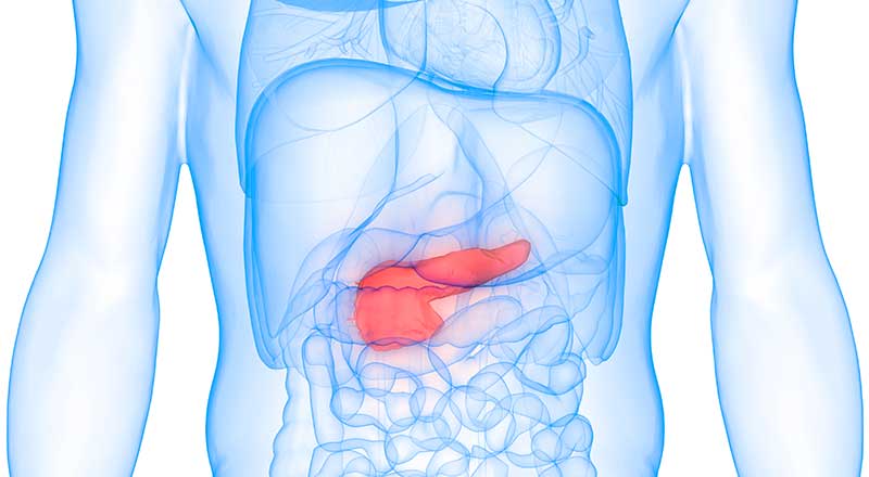
Located behind the stomach, the pancreas is a long, flat gland responsible for producing enzymes that help with blood sugar regulation and digestion.
If you have pancreatic disease (pancreatitis), this gland becomes inflamed. One approach to diagnosis and treatment a GI doctor may recommend if it’s suspected you have this condition is endoscopic retrograde cholangiopancreatography (ERCP).
What is ERCP?
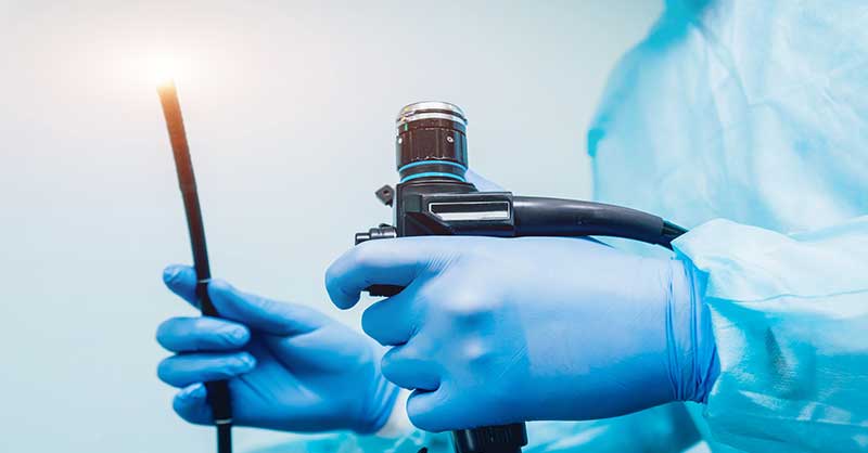
Endoscopic retrograde cholangiopancreatography is a procedure that can be done for either diagnostic or treatment purposes. It combines the use of X-rays with the use of a long, flexible tube with a light source and camera attached called an endoscope. Inserted through the mouth, the scope is used to view the stomach and part of the small intestine. The pancreas can also be viewed and accessed with the scope.


How Do You Prepare for an ERCP?
Let the GI doctor know if you’ve had previous reactions to contrast dye, which is used during the procedure, or if you’re pregnant. Typically, patients are asked not to eat or drink liquids for at least 8 hours prior to having the procedure. Antibiotics may be given to you before the procedure if you have heart valve disease. An ERCP is a same-day procedure usually done with some type of anesthesia and/or sedation. Patients are often awake during the procedure.
What Happens During the Procedure?
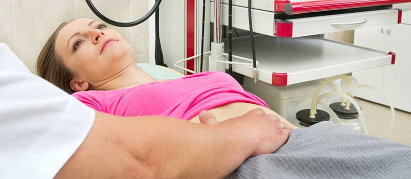
During an ERCP, you will be positioned on an X-ray table in a way that allows the endoscope to be comfortably inserted via your mouth. If you need sedation, it’s usually delivered through an intravenous (IV) line. Numbing medicine is used to prevent gagging and keep you from swallowing as the procedure is performed. However, your saliva will be suctioned out of your mouth.
The endoscope is inserted into an area known as the biliary tree, a system of vessels that allows secretions to travel from the pancreas and other structures in the same area to other parts of the body by way of a series of ducts. A contrast dye is inserted into the ducts through a tube that’s fed through the endoscope. A series of X-rays will then be taken. You may be asked to shift your position when this happens.
The tube for the contrast dye will be repositioned after the X-rays are taken so dye can be injected into the pancreatic duct. X-rays are once again taken. Tissue or fluid samples are sometimes collected as well. If a blockage is detected that’s affecting the pancreas, it may be removed while the endoscope is still in place. It also may be possible to make other repairs to affected ducts with endoscopic techniques.
What Happens After an ERCP?
In some cases, pancreas surgery is scheduled for another time if it’s discovered that diseased tissue needs to be removed and it’s not possible to do so with the endoscope. One option is what’s known as the Whipple procedure (pancreaticoduodenectomy). When the ERCP procedure is over, you’ll be able to eat and drink again once your gag reflexes return.
Controlling underlying conditions like diabetes, not smoking, seeking immediate treatment for gallstones, and avoiding excessive alcohol consumption are some of the ways you may be able to prevent pancreatic disease from developing. You may also be at risk for developing problems with your pancreas if you have cystic fibrosis or a family history of pancreatitis.

